40 label the structures found within a skeletal muscle.
PLAQUENIL® - Food and Drug Administration Neuromuscular Reactions: Skeletal muscle palsies or skeletal muscle myopathy or neuromyopathy leading to progressive weakness and atrophy of proximal muscle groups which may be associated with mild sensory changes, depression of tendon reflexes and abnormal nerve conduction. Ocular Reactions: Antipsychotic drugs in pregnancy: a review of their maternal … Risk of structural teratogenicity. All antipsychotics cross the placenta [Newport et al. 2007] and, as such, consideration needs to be given to their potential to cause structural or functional dysgenesis of fetal organs and/or skeletal structures when exposure occurs in first trimester.It is generally considered that the baseline population rate for malformation in the general …
Solved Label the structures found within a skeletal muscle. - Chegg Label the structures found within a skeletal muscle. Fascicle Sarcoplasmic reticulum Muscle fiber Nerve Filaments Sarcolemma Blood vessels Myofibril Reset Question: Label the structures found within a skeletal muscle. Fascicle Sarcoplasmic reticulum Muscle fiber Nerve Filaments Sarcolemma Blood vessels Myofibril Reset This problem has been solved!

Label the structures found within a skeletal muscle.
Anatomy, Skeletal Muscle - StatPearls - NCBI Bookshelf The individual bundles of muscle fibers in a skeletal muscle are known as fasciculi. The outermost connective tissue sheath surrounding the entire muscle is known as epimysium. The connective tissue sheath covering each fasciculus is known as perimysium, and the innermost sheath surrounding individual muscle fiber is known as endomysium.[9] Skeletal muscle mass and distribution in 468 men and women … 22/02/2020 · We employed a whole body magnetic resonance imaging protocol to examine the influence of age, gender, body weight, and height on skeletal muscle (SM) mass and distribution in a large and heterogeneous sample of 468 men and women. Men had significantly (P < 0.001) more SM in comparison to women in both absolute terms (33.0 vs. 21.0 kg) and relative to … SKELETAL MUSCLE ORGANIZATION During muscle contraction, the myosin heads link the thick and thin myofilaments together, forming cross bridges that cause the thick and thin myofilaments to slide over each other, resulting in shortening of each sarcomere, each skeletal muscle fiber, and the muscle as a whole-much like the two parts of an extension ladder slide over each other. To summarize, in …
Label the structures found within a skeletal muscle.. Solved Label the structures found within a skeletal muscle. | Chegg.com Expert Answer 100% (3 ratings) Tendon Fascia Epimysium Perimysiu … View the full answer Transcribed image text: Label the structures found within a skeletal muscle. Muscle fiber Myofibril Sarcoplasmic reticulum Filaments Perimysium Sarcolemma Epimysium Endomysium Fascia Epimysium Endomysium Fascia Tendon Fascicle Reset Nervous System: Explore the Nerves with Interactive Anatomy 02/11/2020 · Neurons, also known as nerve cells, communicate within the body by transmitting electrochemical signals. Neurons look quite different from other cells in the body due to the many long cellular processes that extend from their central cell body. The cell body is the roughly round part of a neuron that contains the nucleus, mitochondria, and most of the cellular organelles. … Label structure of skeletal muscle Diagram | Quizlet a long, filamentous organelle found within muscle cells that has a banded appearance tendon cordlike extension of connective tissue beyond the muscle, serving to attach it to the bone sarcolemma plasma membrane of a muscle cell epimysium just deep to the deep fascia (surrounds entire muscle) perimysium Skeletal Muscle Organization: Connective Tissue and Layers Muscles are attached to bones by tendons Our bodies contain numerous skeletal muscle organs - for example, the biceps brachii of the arm and the gastrocnemius of the leg. The muscles that move and...
A&P 139 Digestive System Flashcards - Quizlet Label the major regions of the stomach and its associated structures in this frontal view. abdominal part of esophagus Complete each sentence describing the blood flow to and from the liver. welcome to Ms. stephens' anatomy and Physiology and … Unit 5: Muscular System Student Learning Goals: I can identify smooth, skeletal, and cardiac muscle tissue under a microscope and state the function of each.; I can identify the component parts of a muscle: fascicle, myofibril, fiber, nucleus of cell, body of muscle.; I can identify the major muscles of the human body.; I can analyze experimental data using the Moving Arm … Chapter 9 Muscular Homework Flashcards - Quizlet Therefore, layers of _______ ________ enclose and separate all parts of a skeletal muscle connective tissue Label the structures found within a skeletal muscle. Place in order from largest to smallest the components of a skeletal muscle. Label the components of a myofibril. Label the myofibril and its components. Skeletal muscle mass and distribution in 468 men and women ... Feb 22, 2020 · We employed a whole body magnetic resonance imaging protocol to examine the influence of age, gender, body weight, and height on skeletal muscle (SM) mass and distribution in a large and heterogeneous sample of 468 men and women. Men had significantly (P < 0.001) more SM in comparison to women in both absolute terms (33.0 vs. 21.0 kg) and relative to body mass (38.4 vs. 30.6%). The gender ...
Skeletal Muscle - Anatomy & Physiology - UH Pressbooks Skeletal Muscle Fibers. Because skeletal muscle cells are long and cylindrical, they are commonly referred to as muscle fibers. Skeletal muscle fibers can be quite large for human cells, with diameters up to 100 μm and lengths up to 30 cm (11.8 in) in the Sartorius of the upper leg.During early development, embryonic myoblasts, each with its own nucleus, fuse with up to hundreds of other ... SKELETAL MUSCLE ORGANIZATION Each skeletal muscle cell, also called a muscle fiber, develops as many embryonic myocytes fused into one long, multi-nucleated skeletal muscle cell. These muscle fibers are bound together into bundles, or fascicles, and are supplied with a rich network of blood vessels and nerves. The fascicles are then bundled together to form the intact muscle. Skeletal Muscle Tissue Quiz for Anatomy - Registered Nurse RN The answer is b, epimysium. This connective tissue layer surrounds the entire muscle organ. The prefix epi means upon, just like the epidermis is the outermost later upon the skin. 2. A bundle of several skeletal muscle fibers, as pictured in figure 2, is called: a. Sarcomere. b. Myofibril. Skeletal Muscle: Definition, Function, Structure, Location | Biology ... Skeletal muscle, as the name implies, is any muscles that connects to and controls the motions of the skeleton. In all there are somewhere between 600 and 900 muscles in the human body, but an exact number is hard. Many muscles are obscurely small or are sometimes grouped together with similar muscles.
9 best epithelial tissue images on Pinterest | Nursing board, Anatomy ...
Skeletal muscle: A review of molecular structure and function, in ... 1. INTRODUCTION. Striated muscle is composed of two major muscle types—skeletal and cardiac. While the cardiac (heart) muscle functionally represents a set of self‐stimulating, non‐fatiguing muscle cells with an intermediate energy requirement, skeletal muscle represents a set of innervated, voluntary muscle cells that exhibit fatigue with high energy requirements (e.g., muscles of the ...

Muscle Tissue Diagram Labeled / Human Physiology Muscle : Start ...
Skeletal Muscle | Anatomy and Physiology | | Course Hero Inside each skeletal muscle, muscle fibers are organized into individual bundles, each called a fascicle, by a middle layer of connective tissue called the perimysium.This fascicular organization is common in muscles of the limbs; it allows the nervous system to trigger a specific movement of a muscle by activating a subset of muscle fibers within a bundle, or fascicle of the muscle.
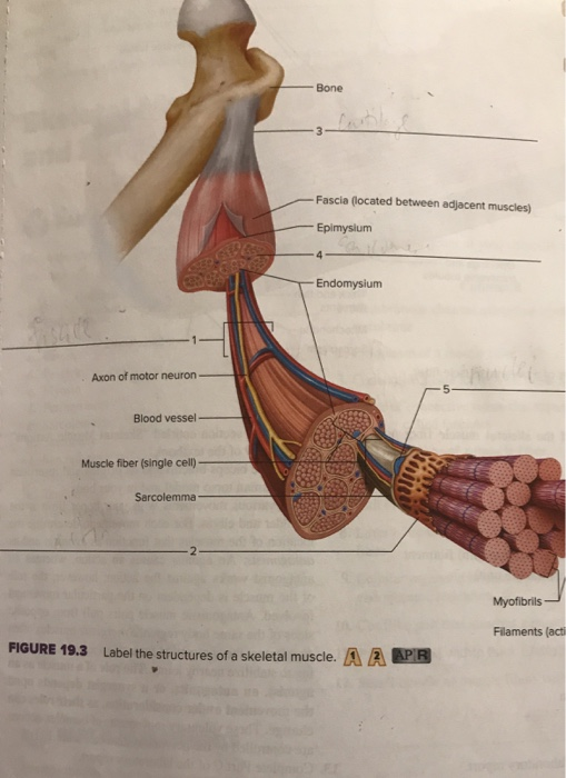
Solved: Bone Fascia (located Between Adjacent Muscles) Epi... | Chegg.com
Skeletal Muscle Tissue Anatomy and Structure - Registered Nurse RN Skeletal Muscle Structure Each skeletal muscle is considered an organ, and it's made up of connective tissue layers, muscle fibers, blood vessels, and nerves. Skeletal muscles attach to the bones through tendons or through a direct attachment. As you look at this muscle diagram, you'll notice an outer layer of connective tissue called epimysium.

Human Physiology – Introduction To The Muscular System | Genius
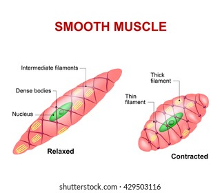
welcome to Ms. stephens' anatomy and Physiology and ... Unit 5: Muscular System Student Learning Goals: I can identify smooth, skeletal, and cardiac muscle tissue under a microscope and state the function of each.; I can identify the component parts of a muscle: fascicle, myofibril, fiber, nucleus of cell, body of muscle.
Muscle Tissue Diagram Labeled / Human Physiology Muscle : Start ...
A&P chapter 9 Flashcards - Quizlet Place in order, from largest to smallest, the components of a skeletal muscle muscle, fascicle, muscle fiber, myofibril, myofilaments Label the parts of a lever system Which of the following is not true Red fibers have fewer mitochondria than white. Acetylcholine (ACh) is released from motor neurons and enters the synaptic cleft
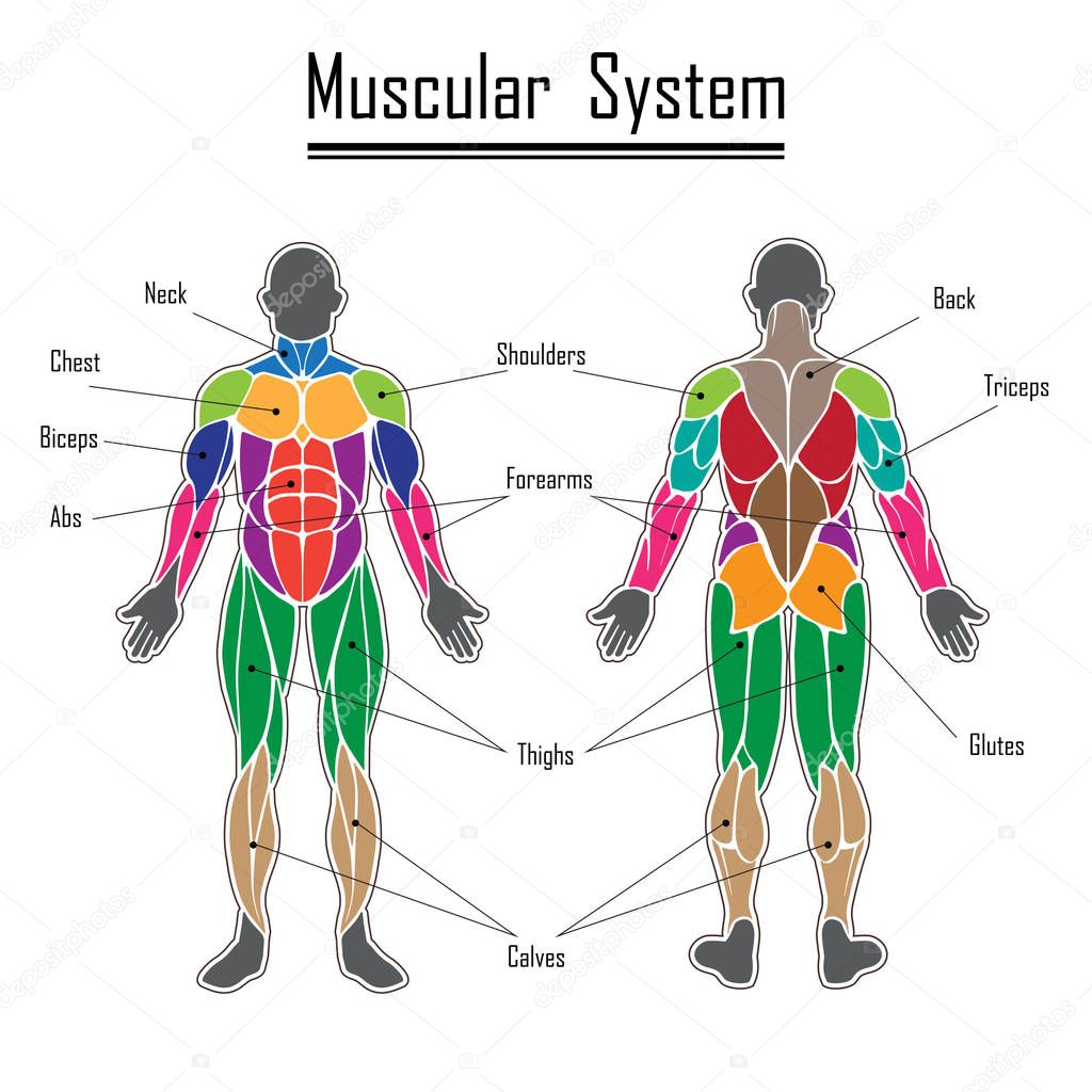
Diagram Of All Muscles In Body / muscle diagram - Free Large Images ...
The excitation–contraction coupling mechanism in skeletal muscle Jan 24, 2014 · The most suitable dyes to study ECC in skeletal muscle seem to be the low affinity Ca 2+ dyes, Mag-Fura-2 and Mag-Fluo-4 (Fig. 1), since they are well known and can reliably track fast, large and brief Ca 2+ transients such as those found in skeletal muscle (Hollingworth et al. 2009; Baylor and Hollingworth 2011; Calderón et al. 2010, 2013).

Image result for skeletal muscle cells diagram with mitochondria ...
Solved Label the structures found within a skeletal muscle. - Chegg Question: Label the structures found within a skeletal muscle. Fascicle Filaments Perimysium Myofibril Epimysium Muscle fiber Fascia Sarcolemma Tendon Endomysium Reset This problem has been solved! See the answer Label the structures found within a skeletal muscle. Show transcribed image text Expert Answer
Post a Comment for "40 label the structures found within a skeletal muscle."