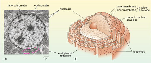
42 label the transmission electron micrograph of the nucleus.
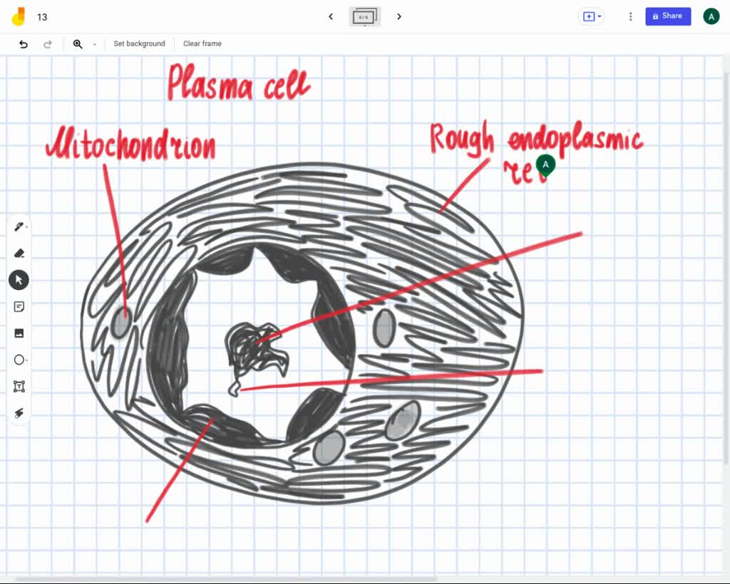
Transmission electron micrograph of cell nucleus - Stock Image - G455 ... Cell nucleus. Transmission electron micrograph of the nucleus (circular) of a mouse liver cell. Surrounding the nucleus is delicate nuclear membrane, which contains gaps called nuclear pores that allow large molecules to pass out into the cell cytoplasm. The dark area in the lower part of the nucleus is the nucleolus. Transmission Electron Microscope (TEM) - Uses, Advantages and Disadvantages A Transmission Electron Microscope produces images via the interaction of electrons with a sample. TEMs are costly, large, cumbersome instruments that require special housing and maintenance. They are also the most powerful microscopic tool available to-date, capable of producing high-resolution, detailed images 1 nanometer in size. ...
The Transmission Electron Microscope | CCBER - UC Santa Barbara Transmission electron microscopes (TEM) are microscopes that use a particle beam of electrons to visualize specimens and generate a highly-magnified image. TEMs can magnify objects up to 2 million times. In order to get a better idea of just how small that is, think of how small a cell is.

Label the transmission electron micrograph of the nucleus.
Transmission Electron Microscope (With Diagram) - Biology Discussion Transmission Electron Microscope (With Diagram) In this article we will discuss about the design of transmission electron microscope, explained with the help of a diagram. In TEM a finely focused beam of electrons from an electron gun is passed through a specially prepared ultra thin section of the specimen. The beam is focused on a small area ... histology.medicine.umich.edu › resourcesPeripheral Nervous System | histology - University of Michigan Also, note the nucleus and cytoplasmic organelles of the Schwann cell. Remember that the myelin is part of the Schwann cell, not of the axon. 55 Peripheral nerve - Unmyelinated Nerve Fibers - Cross Section View Virtual EM Slide Unmyelinated Nerve Fibers (cross section). The axons seen in this electron micrograph are all non-myelinated. Transmission electron micrograph (TEM) identifying immunogold labeled ... Transmission electron micrograph (TEM) identifying immunogold labeled ESR1 and caveolin-1 proteins in the uterine artery endothelial cells derived from the pregnant state (P-UAEC). (A) IgG...
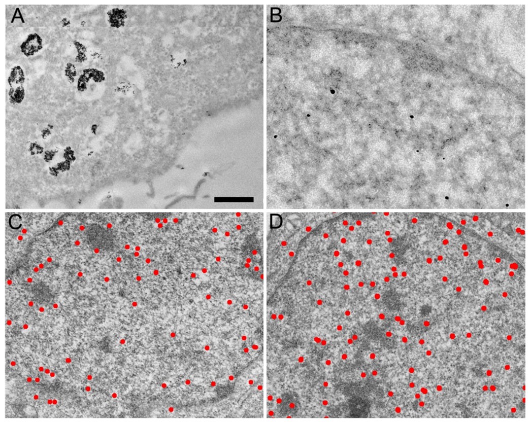
Label the transmission electron micrograph of the nucleus.. anatomy 10.png - Label the transmission electron micrograph... document. 14. operations which are not found in 4 One is an auxiliary inversion operation by. document. 258. Based on the results of internal analysis organization may develop strategies. document. 73. A CPU bound program will typically have a a few very short CPU bursts b many. Transmission Electron Micrograph of transfected HL-1 cells labeled for ... Transmission Electron Micrograph of transfected HL-1 cells labeled for TMEM43 with immunogold. A and B. Single immunogold labeling experiments used 15 nm gold particles to label GFP. A.... Label This Transmission Electron Micrograph Of A Relaxed ... - Blogger Label this transmission electron micrograph of relaxed sarcomeres by clicking and dragging the labels to the correct location . Label the following image using the terms provided. Note how the sarcomeres are extended to only approximately 120 % . IMG_2132 - FIGURES Label this transmission electron from Chapter 14 & 15 Flashcards Flashcards | Quizlet Label the transmission electron micrograph based on the hints provided. Place the following tonsils in order based on their location from superior to inferior. Label the structures in the photomicrograph based on the hints provided
Label This Transmission Electron Micrograph : The Corresponds To The ... Label the transmission electron micrograph of the. Transmission electron microscopy (tem) is a microscopy technique in which a beam of electrons is transmitted through a specimen to form an image. Interpretation of electron micrographs to identify organelles and deduce the functions of specialized cells. No microtubule labeling is evident. Label the transmission electron micrograph of the cell. 0 Nucleus ... Label the transmission electron micrograph of the cell. 0 Nucleus rences Mitochondrion Heterochromatin Peroxisome Vesicle ULAR bumit Click and drag each label into the correct category to indicate whether it pertains to the cytoplasm or the plasma membrane. Solved Label the transmission electron micrograph of the - Chegg Expert Answer. 100% (23 ratings) Transcribed image text: Label the transmission electron micrograph of the nucleus. Nuclear envelope Nucleolus Nucleus Heterochromatin Reset Zoom. Label the transmission electron micrograph of the nucleus. - Transtutors Copy And Paste 5 Micrographs With Magnifications That Fall Within The Specified Ranges Into The Text Answer Box. Be Sure To Label Your Images With The Appropriate Name And Magnification. Post Them In The Specified Order. 1. Transmission... Posted 6 months ago Recent Questions in Economics - Others Q:
en.wikipedia.org › wiki › RotavirusRotavirus - Wikipedia Electron micrograph of a rotavirus infected enterocyte (top) compared to an uninfected cell (bottom). The bar = approx. 500 nm Rotaviruses replicate mainly in the gut , [74] and infect enterocytes of the villi of the small intestine , leading to structural and functional changes of the epithelium . [75] Cell Nucleus - function, structure, and under a microscope The nucleus is a double-layer membrane organelle. It consists of the nuclear envelope, DNA (chromatin), nucleolus, nucleoplasm, and the nuclear matrix. The main function of the nucleus is to control cell activities and carry genetic information to pass to the next generation. A eukaryotic cell typically has only one nucleus. Eukaryotic Cells | Biology I - Lumen Learning The centrosome is a region near the nucleus of animal cells that functions as a microtubule-organizing center. It contains a pair of centrioles, two structures that lie perpendicular to each other. ... This transmission electron micrograph shows a mitochondrion as viewed with an electron microscope. Notice the inner and outer membranes, the ... PDF Identifying Organelles from an Electron Micrograph The electron micrograph displayed below illustrates many of the plant cell characteristics discussed The cell wall, large central vacuole and chloroplasts are clearly visible Also visible is the clearly defined nucleus containing chromatin Nucleus Chromatin The vacuole in this mature plant cell from a leaf is large, and occupies about 80% of
A tour of the cell: View as single page - Open University Figure 2 (a) A transmission electron microscope. (b) A transmission electron micrograph of a frog leukocyte (white blood cell). The nucleus and nucleolus (Section 4.3), mitochondria (Section 4.10) and Golgi apparatus (Section 4.7) can be seen. The dark area of the nucleus contains densely packed DNA.
((a) Draw and label a diagram to show the structure of a nucleus, as ... Click here👆to get an answer to your question ️ ((a) Draw and label a diagram to show the structure of a nucleus, as seen using a transmission electron microscope.
Electron Micrographs of Cell Organelles | Zoology - Biology Discussion This is an electron micrograph of nucleus. (Fig. 17 & 18): (1) Nucleus was discovered by Brown (1831). (2) It is a characteristic entity of almost all eukaryotic cells except mammalian RBCs. (3) The nucleus is generally one but may also be two, four or many.
› createJoin LiveJournal Password requirements: 6 to 30 characters long; ASCII characters only (characters found on a standard US keyboard); must contain at least 4 different symbols;
Electron Micrographs - University of Oklahoma Health Sciences Center You should concentrate on the similarities in form that permit identification of the components irrespective of cell type. Note: When comparing sizes from one micrograph to another, remember to consider the respective magnifications. Figure 1 Micrograph of a nucleus. 1. Heterochromatin 2. Euchromatin 3. Nucleolus 4. Nucleolar associated chromatin
Nucleus - Electron Micrograph - University of Tulsa Slide 5 of 36
Transmission electron micrograph of a cell nucleus Coloured transmission electron micrograph of the nucleus of a pancreatic cell, surrounded by cytoplasm. Every cell nucleus contains a complete copy of all the organism's genes, which are encoded in DNA (deoxyribonucleic acid) molecules in the chromosomes. In human cell nuclei there are 46 chromosomes - 23 inherited from the mother and 23 from ...
Label This Transmission Electron Micrograph - Kaiden Brown Label the transmission electron micrograph of the nucleus. Label the transmission electron micrograph of the nucleus. Transmission electron microscopy (tem) is a microscopy technique in which a beam of electrons is transmitted through a specimen to form an image. Figures label this transmission electron micrograph ( 16, . CIN2003. Ian Roberts.
Solved Label the transmission electron micrograph of the - Chegg Question: Label the transmission electron micrograph of the cell. 0 Nucleus rences Mitochondrion Heterochromatin Peroxisome Vesicle ULAR bumit Click and drag each label into the correct category to indicate whether it pertains to the cytoplasm or the plasma membrane.
95 Electron Micrograph Nucleus Premium High Res Photos - Getty Images Find Electron Micrograph Nucleus stock photos and editorial news pictures from Getty Images. Select from premium Electron Micrograph Nucleus of the highest quality. ... carcinoma cell, colored transmission electron micrograph (tem) - electron micrograph nucleus stock illustrations. whitefish mitosis - interphase. embryo (blastula). shows ...
Neuron under Microscope with Labeled Diagram - AnatomyLearner The nucleus is the spherical or elliptical structure in the neuron containing euchromatic staining (pale staining). Again, the shape of the nucleus of a neuron is generally large because of the little cell body cytoplasm. There is a prominent nucleolus evident in the nucleus of a neuron.
Looking at the Structure of Cells in the Microscope Determining the detailed structure of the membranes and organelles in cells requires the higher resolution attainable in a transmission electron microscope. Specific macromolecules can be localized with colloidal gold linked to antibodies. Three-dimensional views of the surfaces of cells and tissues are obtained by scanning electron microscopy.
› 43392556 › Cambridge_IGCSECambridge IGCSE Biology Third Edition Hodder Education Enter the email address you signed up with and we'll email you a reset link.
quizlet.com › 561647498 › labeling-the-cell-flash-cardsLabeling the Cell Flashcards | Quizlet Label the transmission electron micrograph of the nucleus. membrane bound organelles golgi apparatus, mitochondrion, lysosome, peroxisome, rough endoplasmic reticulum nonmembrane bound organelles ribosomes, centrosome, proteasomes cytoskeleton includes microfilaments, intermediate filaments, microtubules Identify the highlighted structures
› books › NBK21052Glossary - Molecular Biology of the Cell - NCBI Bookshelf micrograph. Photograph of an image seen through a microscope. May be either a light micrograph or an electron micrograph depending on the type of microscope employed. microinjection. Injection of molecules into a cell using a micropipette. micron (m or micrometer) Unit of measurement often applied to cells and organelles.
docer.tips › solomon-berg-martin-biology-9thSolomon, Berg, Martin - Biology 9th Edition - PDF Free Download H1N1, the virus that causes H1N1 influenza (flu). H1N1 virus particles (blue) are visible on a cell (green). When this virus emerged, the human immune system was unfamiliar with its new combination of genes. As a result, the virus spread easily, causing a pandemic. The scanning electron micrograph (SEM) has been color-enhanced. T
Transmission electron micrograph (TEM) identifying immunogold labeled ... Transmission electron micrograph (TEM) identifying immunogold labeled ESR1 and caveolin-1 proteins in the uterine artery endothelial cells derived from the pregnant state (P-UAEC). (A) IgG...
histology.medicine.umich.edu › resourcesPeripheral Nervous System | histology - University of Michigan Also, note the nucleus and cytoplasmic organelles of the Schwann cell. Remember that the myelin is part of the Schwann cell, not of the axon. 55 Peripheral nerve - Unmyelinated Nerve Fibers - Cross Section View Virtual EM Slide Unmyelinated Nerve Fibers (cross section). The axons seen in this electron micrograph are all non-myelinated.
Transmission Electron Microscope (With Diagram) - Biology Discussion Transmission Electron Microscope (With Diagram) In this article we will discuss about the design of transmission electron microscope, explained with the help of a diagram. In TEM a finely focused beam of electrons from an electron gun is passed through a specially prepared ultra thin section of the specimen. The beam is focused on a small area ...














Post a Comment for "42 label the transmission electron micrograph of the nucleus."