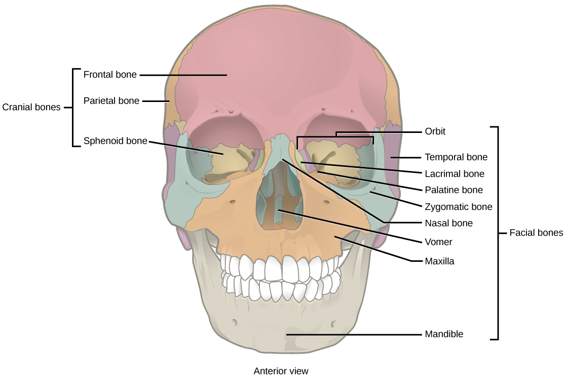
41 figure 14.1 label the anterior bones and features of the skull
Snake - Wikipedia As a result, the vertebrae anterior to the hindlimb buds (when present) all have the same thoracic-like identity (except from the atlas, axis, and 1-3 neck vertebrae). In other words, most of a snake's skeleton is an extremely extended thorax. Ribs are found exclusively on the thoracic vertebrae. Multi-tract multi-symptom relationships in pediatric concussion This manuscript aims to address an important issue in the study of concussion: both the brain damage caused by concussion, as well as the behavioral symptoms that result vary widely across individuals. The study uses novel and interesting methods to relate multi-variate diffusion MRI data with multi-variate symptom-related data.
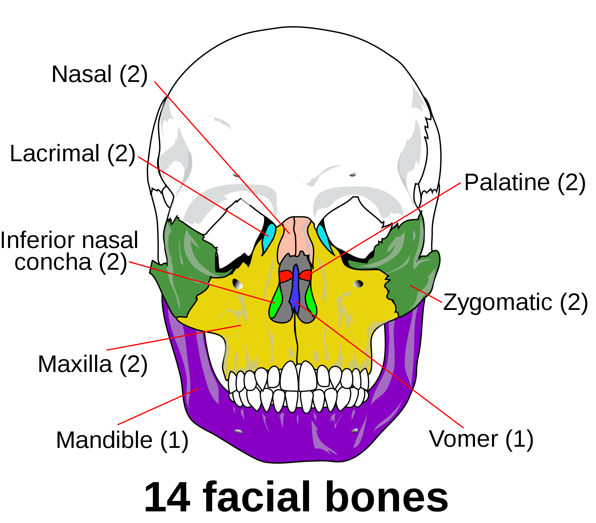
Skull: Anatomy, structure, bones, quizzes | Kenhub The human skull consists of 22 bones (or 29, including the inner ear bones and hyoid bone) which are mostly connected together by ossified joints, so called sutures.The skull is divided into the braincase (neuro cranium) and the facial skeleton (viscerocranium).Its main task is the protection of the most important organ in the human body: the brain.
Figure 14.1 label the anterior bones and features of the skull
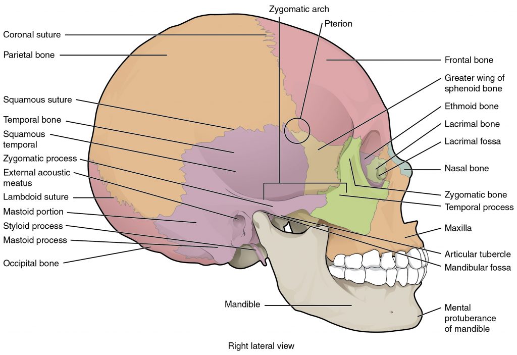
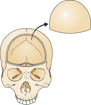
Bones of the Skull | Skull Osteology | Anatomy | Geeky Medics The calvarium, also known as the roof or skull cap, consists of three bones: Frontal bones. Parietal bones. Occipital bones. These bones protect the brain superiorly, but also provide an anchor for important muscles of facial expression and eye movement. The parts of these bones that lie inferior to the brain are considered to be a part of the ... Skull anatomy: Anterior and lateral views of the skull | Kenhub Anterior and lateral views of the skull. The human skull consists of about 22 to 30 single bones which are mostly connected together by ossified joints, so called sutures. The skull is divided into the braincase ( cerebral cranium) and the face ( visceral cranium ). The main task of the skull is the protection of the most important organ in the ... ICD-9-CM Volume 3 - Wikipedia ( 14.1) Diagnostic procedures on retina, choroid, vitreous, and posterior chamber ( 14.2) Destruction of lesion of retina and choroid ( 14.3) Repair of retinal tear ( 14.4) Repair of retinal detachment with scleral buckling and implant ( 14.5) Other repair of retinal detachment
Figure 14.1 label the anterior bones and features of the skull. EOF Osteocalcin attenuates oligodendrocyte ... - Science Advances The bone-derived hormone osteocalcin (OCN) is crucial for brain development and neural cognitive functions, yet the exact roles of OCN in central nervous system (CNS) remain elusive. Here, we find that genetic deletion of OCN facilitates oligodendrocyte (OL) differentiation and hypermyelination in the CNS. Skull Anatomy Labeling - Human Anatomy - GUWS Medical Review a textbook section on the skull. 2. As a review activity, label figures 13.1, 13.2, 13 3, 13.4, and 13.5. 3. Examine the cranial bones of the articulated human skull and the sectioned skull. Also observe the corresponding disarticulated bones. ... Label the anterior bones and features of the skull. (If the line lacks the word bone, label ... Randomized trial of intermittent intraputamenal glial cell line-derived ... Figure 2 B. shows the frequency distribution of motor responses in both groups. Post hoc covariate analyses adjusting for demographic and Parkinson's disease characteristics (see post hoc statistical analysis plan) did not identify any specific clinical features producing change in treatment effect on the primary endpoint.
Neural dynamics underlying birdsong practice and performance After applying a topical anaesthetic (0.25% bupivacaine) and making a vertical incision in the skin over the skull, we made ~1-mm craniotomies in the skull at a predetermined distance from the ... ICD-9-CM Volume 3 - Wikipedia ( 14.1) Diagnostic procedures on retina, choroid, vitreous, and posterior chamber ( 14.2) Destruction of lesion of retina and choroid ( 14.3) Repair of retinal tear ( 14.4) Repair of retinal detachment with scleral buckling and implant ( 14.5) Other repair of retinal detachment Skull anatomy: Anterior and lateral views of the skull | Kenhub Anterior and lateral views of the skull. The human skull consists of about 22 to 30 single bones which are mostly connected together by ossified joints, so called sutures. The skull is divided into the braincase ( cerebral cranium) and the face ( visceral cranium ). The main task of the skull is the protection of the most important organ in the ... Bones of the Skull | Skull Osteology | Anatomy | Geeky Medics The calvarium, also known as the roof or skull cap, consists of three bones: Frontal bones. Parietal bones. Occipital bones. These bones protect the brain superiorly, but also provide an anchor for important muscles of facial expression and eye movement. The parts of these bones that lie inferior to the brain are considered to be a part of the ...
Untitled
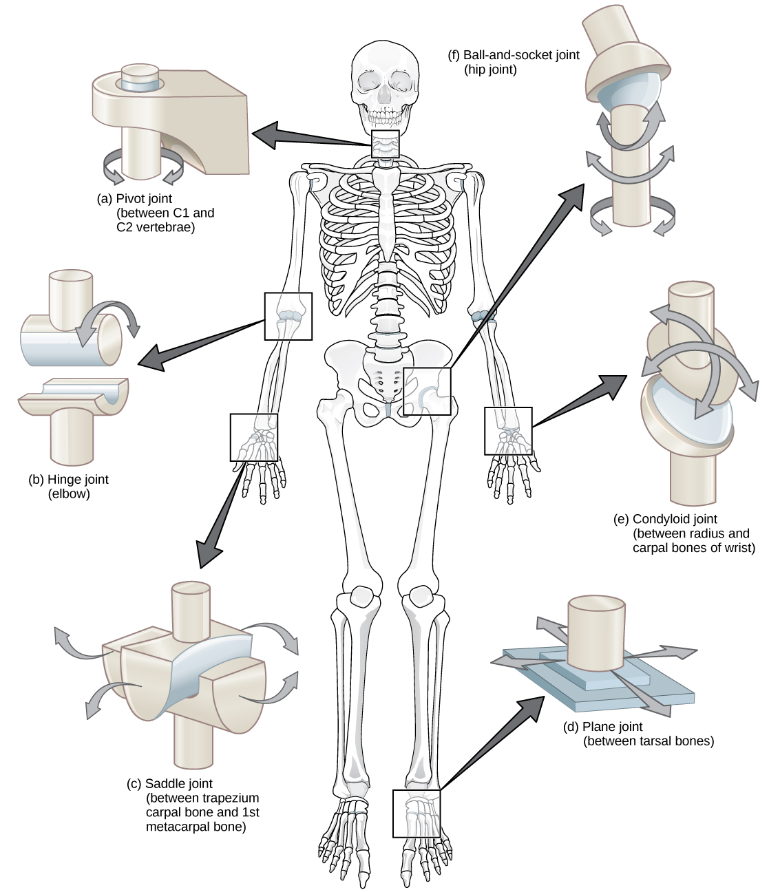
19.3 Joints and Skeletal Movement – Concepts of Biology – 1st ...

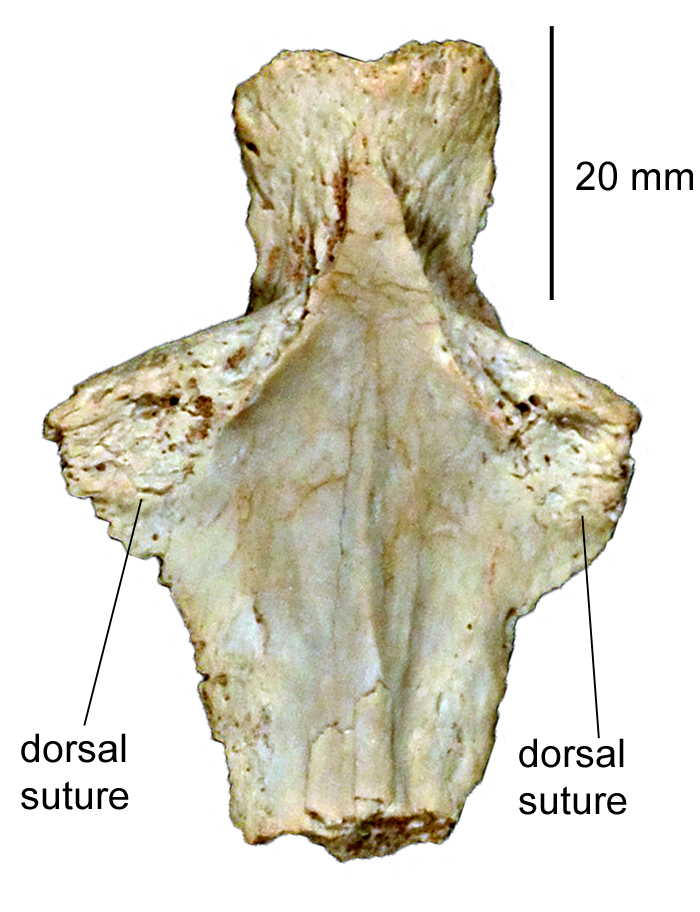
PDF) The mammalian parasphenoid: its cccurrence in marsupials

7.3 The Skull – Anatomy & Physiology

6.2 Bone Classification – Anatomy & Physiology
7.2 Head and Neck Basic Concepts – Nursing Skills
Bones - lawrenceGaltman.com
Human Remains
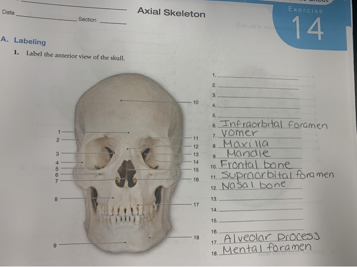
Solved Date Axial Skeleton Exercise Section 14 A. Labeling ...

Assessing Bone Quality of the Spine in Children with ...
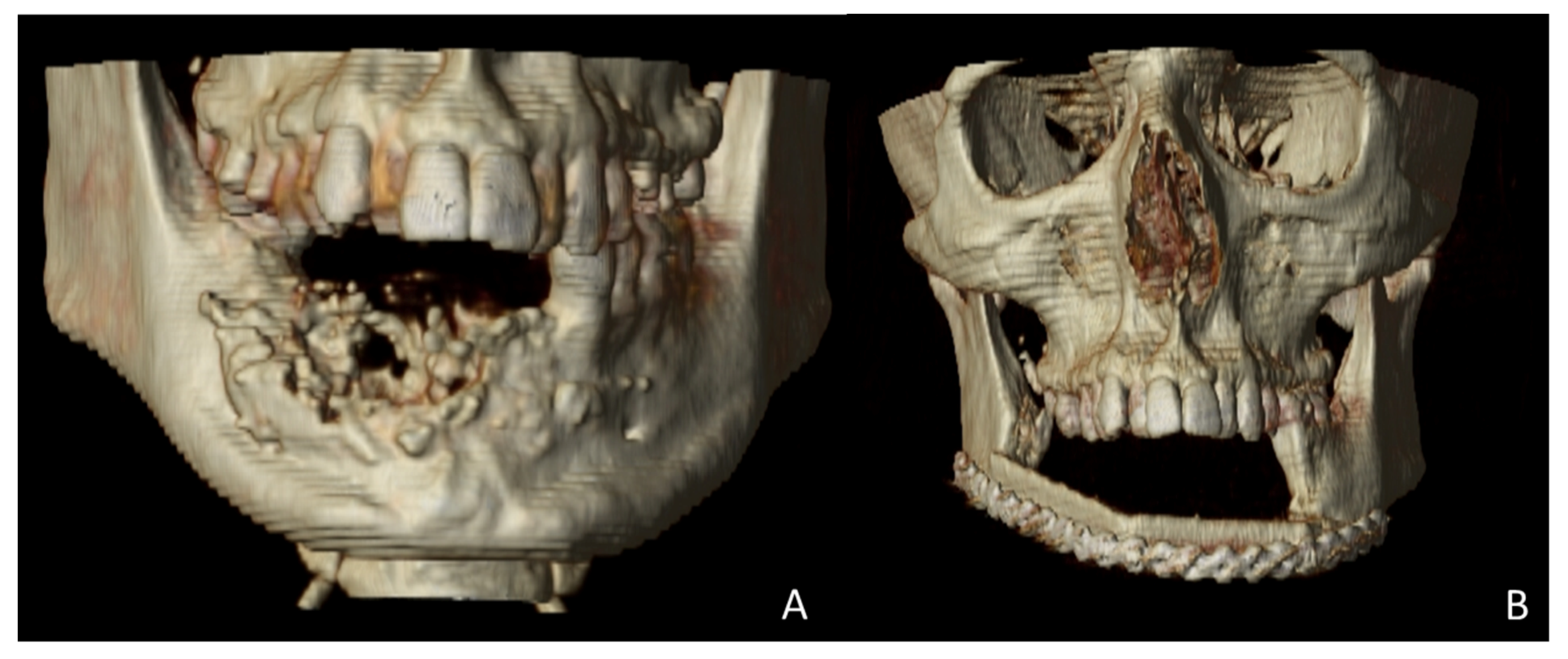
JCM | Free Full-Text | Virtual Surgical Planning ...
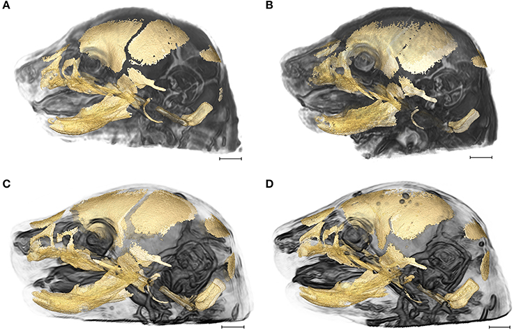
Frontiers | Integration of Brain and Skull in Prenatal Mouse ...
Illustrated Clinical Anatomy, Second Edition
Triassic Turtle Chinlechelys: Figures
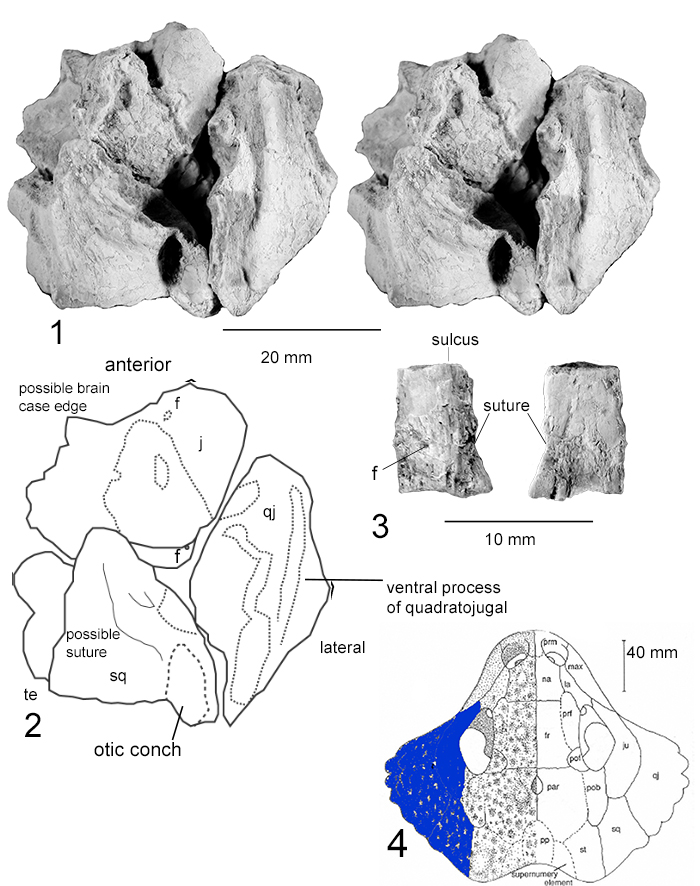
Triassic Turtle Chinlechelys: Figures
Figure 14.1 Label the anterior bones and features of the ...
Figure 14.1 Label the anterior bones and features of the ...

Lab 14: Figure 14.10 Anterior Features of the Skull Diagram ...

A&P 1 Lab Figure 14.1 Bones & Features of the skull (anterior ...

Parasphenoid - an overview | ScienceDirect Topics
Untitled
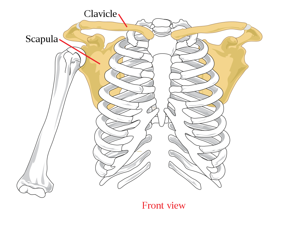
11.3 Divisions of the Skeletal System – Human Biology
11.3 Divisions of the Skeletal System – Human Biology
No Slide Title
7.2 Head and Neck Basic Concepts – Nursing Skills
Open Combined Approaches (Section II) - Integrated Management ...
Figure 14.1 Label the anterior bones and features of the ...

7.3 The Skull – Anatomy & Physiology

7.1 Divisions of the Skeletal System – Anatomy & Physiology

Brachycephalus bufonoides Miranda-Ribeiro 1920 - Plazi ...

BW AP Skull fig 14.1 lab - Figure 14.1 Label the anterior ...
19.1 Types of Skeletal Systems – Concepts of Biology – 1st ...

LAB-14 SKULL.docx - LAB-14 SKULL FIGURE 14.1 1) 2) 3) 4) 5) 6 ...
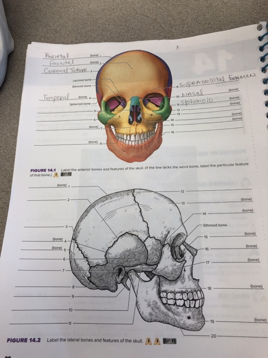
Solved Parietal one)... Frontal Coronel Suture, Labore bo ...
Human Remains

Coronal craniosynostosis influences skull growth and shape in ...
mebooksfree.com
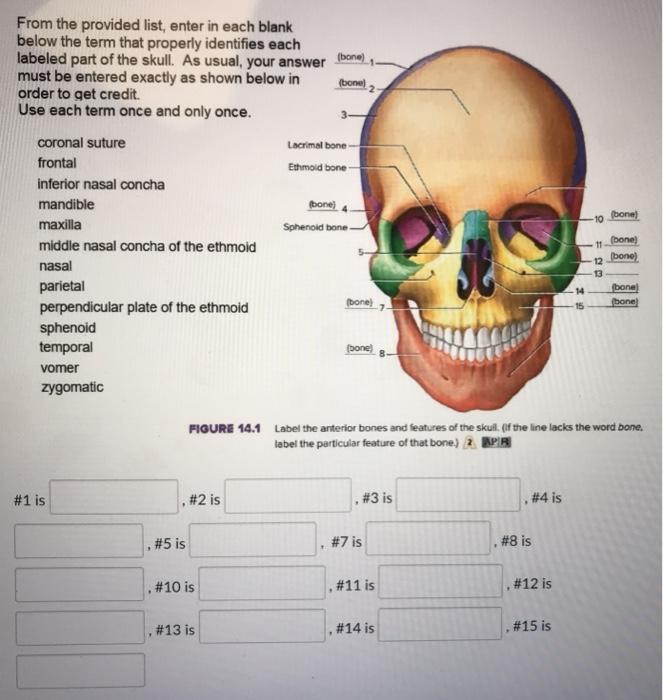
Solved bone) (bone From the provided list, enter in each ...
Figure 14.1 Label the anterior bones and features of the ...

7.3 The Skull – Anatomy & Physiology
Open Combined Approaches (Section II) - Integrated Management ...
Post a Comment for "41 figure 14.1 label the anterior bones and features of the skull"