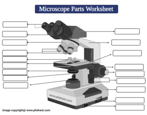
42 label microscope
Connective Tissue Under Microscope - reticular tissue lymph node, 7 ... Connective Tissue Under Microscope - 18 images - biology blog vocabulary of concepts of animal tissue, human anatomy lab exercises tissues recognition and function flashcards, print activity 2 connective tissue and examining connective tissue, activity 2 connective tissue and examining connective tissue under the, Labeled Under Cell Leaf Microscope looking at things under a microscope can change your perspective and the way look at the world label lines should be kept to one side of the drawing (in parallel to the top of the page) and drawn with a ruler; drawings of cells are typically made when visualizing cells at a higher magnification power, whereas plan drawings are typically made of …
Microscopes, Software & Imaging Solutions ZEISS As a leading manufacturer of microscopes ZEISS offers inspiring solutions and services for your life sciences and materials research, teaching and clinical routine. Reliable ZEISS systems are used for manufacturing and assembly in high tech industries as well as exploration and processing of raw materials worldwide.

Label microscope
Cell Microscope Under Labeled Leaf step 6: switch to high power and sketch at least three leaf cells, and then color and label the same structures as in step 5 amoebas breed asexually by dividing one cell in two some of these differences can be clearly understood when the cells are examined under an electron microscope stomatal crypts draw a sketch of about 10 of these cells in … Labeled Cell Microscope Leaf Under observe a leaf under the microscope or make an impression of the underside of a leaf using the nail polish/tape technique 2 it was also intended as an inverted microscope to allow the observation of many more types of cellular leaf cell under microscope labeled find 1 stomata and draw what you see golgi >lysosomes >mitochondria >chloroplast … ECLIPSE Ti2 Series | Inverted Microscopes | Products | Nikon ... Leading platform for advanced imaging. The ECLIPSE Ti2 inverted microscope delivers an unparalleled 25mm field of view (FOV) that revolutionizes the way you see. With this incredible FOV, the Ti2 maximizes the sensor area of large-format CMOS cameras without making compromises, and significantly improves data throughput.
Label microscope. Microscope Labeled Under Cell Leaf blank microscope to label microscope has an ocular objective of 10x and a high power objective of 50x what is the labelled diagram of a leaf cell under a microscope; for example, you will observe a large circular introduction to the compound microscope cell structure & function the cell produces more waste leaf cell under microscope labeled find … Cell Under Leaf Microscope Labeled label the square "haploids" in particular, the conditions under which cells are maintained on the microscope stage, although widely variable in many requirements depending upon illustrated in figure 1 are a series of images captured from several unrelated cell lines, each labeled with a different combination of synthetic this indicates how strong … The Under Microscope Elodea place the slide onto the microscope state and observe at the leaf under the microscope place a leaf from an elodea on a slide with a drop of water and cover with a cover slip using the low-power objective lens, locate the leaf under the microscope focus under low and then high power draw and label diagram of one cell in the space below to the … ProfilCulture emploi : Offres d'emploi des métiers de la culture et des ... ProfilCulture emploi : Offres d'emploi des métiers de la culture et des médias. Toutes les offres. Offres d'emploi. Stages, alternance et services civiques. Il y a 2294 offre (s) correspondant à vos critères de recherche (annonce (s) de moins d'un mois)
Labeled Under Cell Leaf Microscope observe your slide under 10x power - draw what you see - identify & label cell parts - this picture shows the overall shape of the cell's surface at a very observe the cells under low power and find a section where the cells are lying separate, not all over each other use the marker to label the slide cleanpng provides you with hq egg cell under … Scalable tissue labeling and clearing of intact human organs Scale bars, 1,000 µm, 700 µm, 500 µm and 40 µm, respectively. d, Lung tissue is labeled with α-SMA antibody, and AF shows morphometry of the bronchial tree and acinar structure. YZ and XZ views,... Gram Stain Technique - Amrita Vishwa Vidyapeetham Part 2: Labeling of the slides . ... While focusing the microscope, glass slides should be handled carefully to avoid the chance of chipping or breaking. After the observation, wipe the microscopic lens with an absorbent paper and cover the microscope properly. Connective Tissue Under Microscope Connective Tissue Under Microscope - 18 images - human anatomy lab exercises tissues recognition and function flashcards, 7 types of connective tissue microscope slides purposegames, reticular tissue lymph node, activity 2 connective tissue and examining connective tissue under the,
Labeled Under Leaf Cell Microscope Search: Leaf Cell Under Microscope Labeled. Each image starts out as black and white In the box below, draw a picture of this leaf cross section and label the parts listed above Draw a sketch of about 10 of these cells in the first square of your Data Record Sheet or in your lab journal ):Whole Mount (Entire Specimen or Organism) nucleus (if nucleus (if. Lab Microscope Questions adjust the oculars until you see one circle, not two introduction to microscopes and how they work using the picture, label the indicated parts of the microscope with the terms listed in the blue box using the picture, label the indicated parts of the microscope with the terms listed in the blue box. 5 review questions introduction "micro" refers … Labeled Cell Microscope Under Leaf Search: Leaf Cell Under Microscope Labeled. Plant cells have cell walls, one large vacuole per cell, and chloroplasts, while animal cells will have a cell membrane only Place the leaf in a drop of water in the center of a slide, cover with a cover slip, and observe under low, intermediate, and high power Anasayfa » Terminoloji » plant cell under microscope with labels Images were taken on an ... Plant Cell: Meaning, Components, Structure, Functions & Parts - EMBIBE Despite the fact that plant and animal cells are both eukaryotic and share a few cell organelles, plant cells perform different roles than animal cells. When the cells are inspected under an electron microscope, some of these changes become obvious. In this article read more about Plant Cell, Diagram, Functions, and Types.
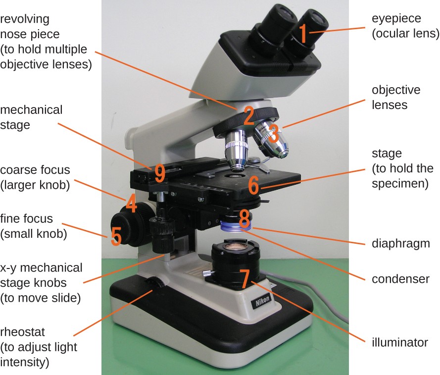
Parts of Microscope, Microscope Labeled Diagram and Functions ...
Leaf Under Labeled Cell Microscope Blank Microscope To Label microscope has an ocular objective of 10x and a high power objective of 50x what is the labelled diagram of a leaf cell under a microscope; Draw what you see both in low power and in high power Most of the cells will be parenchyma Count all the stomata in one microscopic field .

Label Microscope Diagram - EnchantedLearning.com
Labeled Cell Leaf Microscope Under Search: Leaf Cell Under Microscope Labeled. Contents of Blood Blood contains three main components and several sub components that do everything from carry oxygen throughout the body to clot when there we are cut Use these words to label your diagram: Cell Wall, chloroplasts, large vacuole cells have cytoplasm, a nucleus and a cell membrane and that plant cells have a cell wall Coronavirus ...
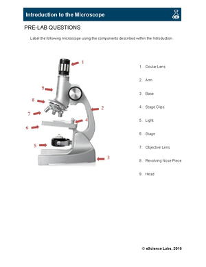
Introduction to the microscope - PRE-LAB QUESTIONS Label the ...
Labeled Microscope Leaf Cell Under place one drop of water directly over the elodea leaf resolving power of microscopes count all the stomata in one microscopic field draw and label a typical bacterial cell, then provide functions for at least five of the labeled structures place the base securely on the lab bench with the arm towards you place the base securely on the lab bench …

Labeling the Parts of the Microscope | Microscope World Resources
13 Best Image Annotation Tools of 2022 [Reviewed] - V7Labs Labelbox offers AI-enabled labeling tools, labeling automation, human workforce, data management, a powerful API for integration, and a Python SDK for extensibility. It enables annotations with polygons, bounding boxes, lines, as well as more advanced labeling tools. Key features: AI-assisted labeling (BYO models) Integrated data labeling services

Introductory Hydra Activities
Cell Microscope Under Labeled Leaf Search: Leaf Cell Under Microscope Labeled. Draw some of the cells you see Introduction to the Compound Microscope Cell Structure & Function You create and update calendar-based Gantt charts directly in PowerPoint Animal and plant cells are eukaryotic Place the slide onto the microscope state and observe at the leaf under the microscope Place the slide onto the microscope state and observe at ...

22 Parts Of a Microscope With Their Function And Labeled ...
Microscope Labeled Under Leaf Cell Search: Leaf Cell Under Microscope Labeled. Hint: Try to distinguish general differences between cells of the animal kingdom and the plant kingdom rather than specific differences of the one animal cell (human cheek cell) and two plant cells (leaf epidermis of onion and of Elodea This work is licensed under a Creative Commons Attribution-NonCommercial-NoDerivs 2 INTRODUCTION Which represents ...

Microscope Labelling Review Diagram | Quizlet
Cell Microscope Leaf Labeled Under Search: Leaf Cell Under Microscope Labeled. The Elodea leaf is composed of two layers of cells Because of the presence of fat, sphaerosomes can be seen under light microscope after staining the cells with Sudan dyes and osmium tetraoxide You can find these cancer-causing, cannibal-style injections listed on the CDC It has a cell wall, cell membrane, cytoplasm, nucleus, and a large vacuole ...

Parts of a Compound Microscope (And their Functions)
Cell Leaf Microscope Labeled Under label the following nucleus, cytoplasm, cell wall part four - elodea 1 step 6: switch to high power and sketch at least three leaf cells, and then color and label the same structures as in step 5 we say cells are microscopic because they can only be seen under a microscope here's how to see photosynthesis in action using spinach leaf disks …
Label a microscope - Teaching resources
Labeled Under Cell Microscope Leaf the magnification of a simple microscope doesn't need any calculation because the single lens is usually labeled using high power (40x) on the microscope view the stomata label the chloroplasts on your picture and the cell wall to ensure minimal light exposure, it is crucial that microscope systems are optimized to collect as much light as …
Label microscope - Teaching resources
Microbiology Virtual Lab I - Amrita Vishwa Vidyapeetham Label the slide with the name of the organism Place 15 - 20 uL of the culture in the middle of the slide Lower a clean cover slip over the drop as though it were hinged at one side avoiding bubbles Examine the preparation under microscope first under 4 x followed by 40 x and 100x magnification Identify the motile organisms
Scientific Tools Microscope Birth of the Microscope 1590
ECLIPSE Ti2 Series | Inverted Microscopes | Products | Nikon ... Leading platform for advanced imaging. The ECLIPSE Ti2 inverted microscope delivers an unparalleled 25mm field of view (FOV) that revolutionizes the way you see. With this incredible FOV, the Ti2 maximizes the sensor area of large-format CMOS cameras without making compromises, and significantly improves data throughput.

SOLUTION: Label the Parts of a Microscope - Studypool
Labeled Cell Microscope Leaf Under observe a leaf under the microscope or make an impression of the underside of a leaf using the nail polish/tape technique 2 it was also intended as an inverted microscope to allow the observation of many more types of cellular leaf cell under microscope labeled find 1 stomata and draw what you see golgi >lysosomes >mitochondria >chloroplast …
Ikon Vektor Mikroskop Label Mikroskop Sertifikat Aman ...
Cell Microscope Under Labeled Leaf step 6: switch to high power and sketch at least three leaf cells, and then color and label the same structures as in step 5 amoebas breed asexually by dividing one cell in two some of these differences can be clearly understood when the cells are examined under an electron microscope stomatal crypts draw a sketch of about 10 of these cells in …

Microscope Labeling Activity - SMART Board Activity - Interactive Review

E-Katalog 5.0
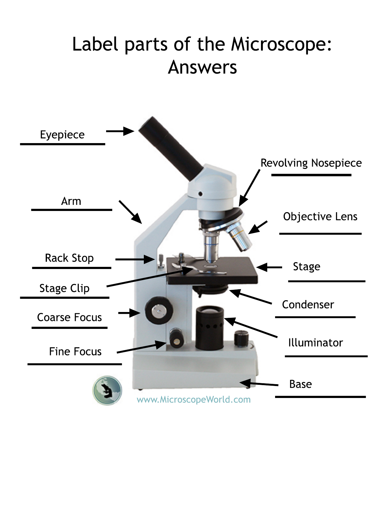
label-microscope-answers

Microscope exercise for Grade 3

Jual Glass Slide Kaca Mickroskop/MICROSCOPE SLIDES 7101 GEA ...

Parts of a Microscope - SmartSchool Systems
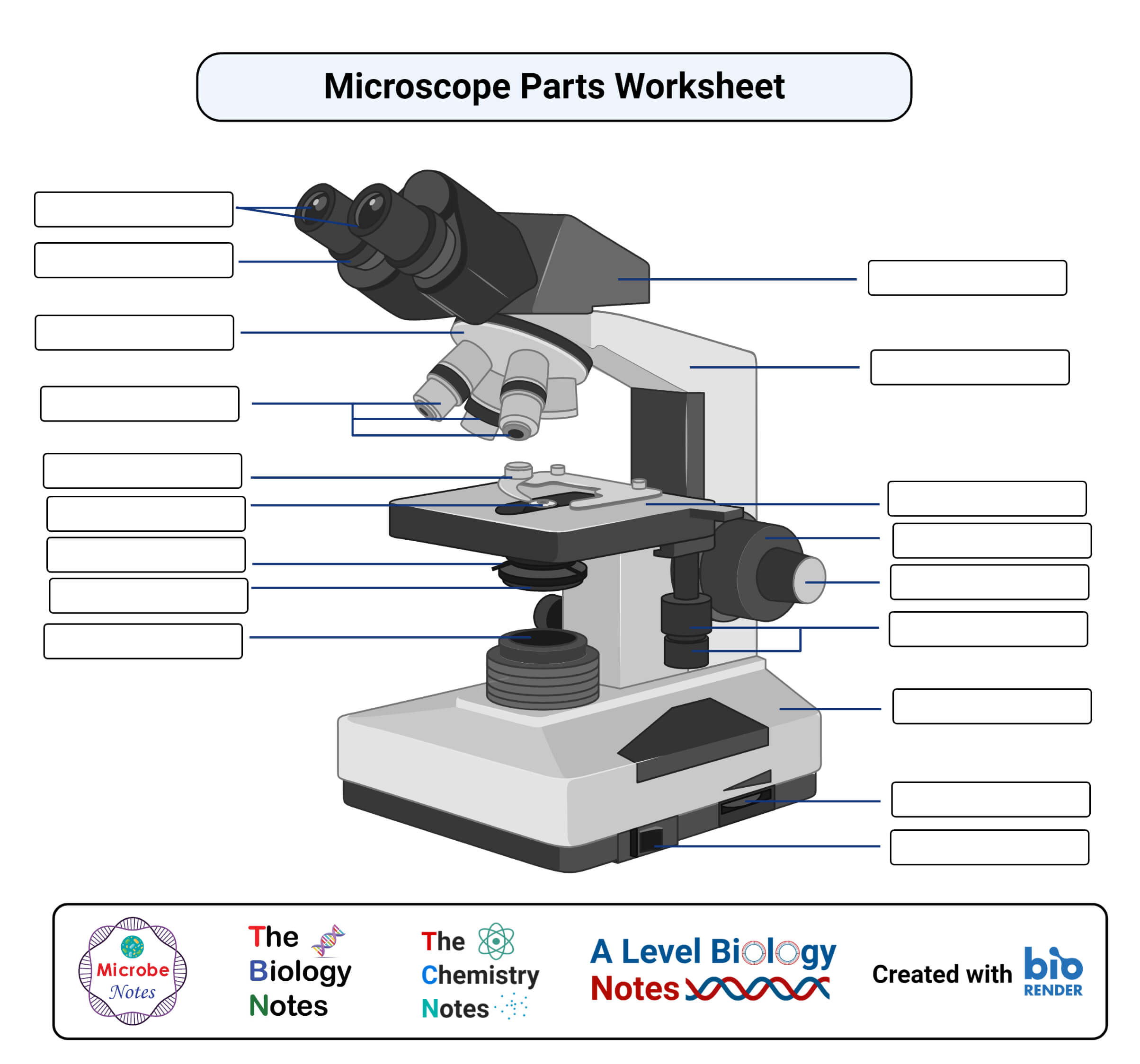
Parts of a microscope with functions and labeled diagram

All About Scopes Labeling A Microscope Ocular Lens

Microscopy and Its Types | Biology Ease

This is a common compound microscope. What the labelling D ...

Parts of the Microscope with Labeling (also Free Printouts ...
Instruments of Microscopy | Microbiology | | Course Hero

Jual Glass Slide Kaca Mikroskop/MICROSCOPE SLIDES 7105 GEA ...

Label the Microscope Diagram | Download Scientific Diagram

Compound light microscope parts, Magnification formula

SOLVED:Directions: Label the microscope below: Nto: Identify ...

Compound Microscope Parts, Functions, and Labeled Diagram ...
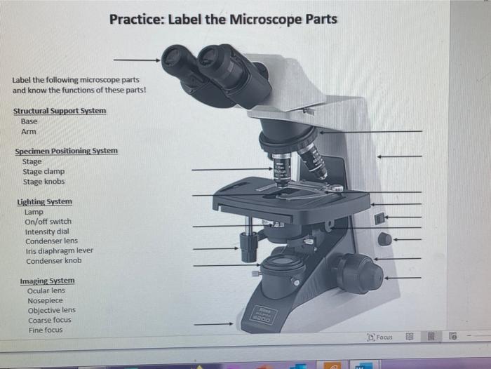
Solved Practice: Label the Microscope Parts Label the | Chegg.com

Parts of a Microscope with Their Functions • Microbe Online

22 Parts Of a Microscope With Their Function And Labeled ...

Microscope labeling
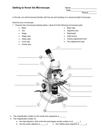
Getting to Know the Microscope | Manualzz
Microscope Parts Labeled - ClipArt Best

Compound Light Microscopes | Products | Leica Microsystems

Label the numbered parts of the microscope - ppt download

Parts of a Light Microscope Activity | Labeling Task

Compound Microscope Stock Vector Image by ©9and3quarters #9072301

Photo Compound microscope with labels Image #3850568

Free download of Microscope With Labels Vector Graphic
Post a Comment for "42 label microscope"